The Time-of-Flight Positron Emission Tomography (TOF-PET) is the most diffuse technology for diagnostic imaging employed in oncology, which allows to reconstruct the image of the tumor tissues with a spatial accuracy of few centimeters. To employ this technology for prevention is necessary to be able to detect embryonal tumors with sizes of few millimeters.
This accuracy, actually not possible, could be reached in the future thanks the new scintillating material realized by the researchers from the University of Milano-Bicocca, as described in the paper “Composite fast scintillators based on high-Z fluorescent metal–organic framework nanocrystals” recently published on Nature Photonics.
The material has been developed by a research team for the Department of Material Science lead by professors Angelo Monguzzi, Anna Vedda e Angiolina Comotti, in collaboration with Dr. Luca Gironi, a researcher from the Department of Physics.
The TOF-PET technique allow to image a tissue properly labelled with a radioactive marker, by exploiting the interaction of the emitted high energy radiation with a scintillator, a material that is able to emit a light pulse as a consequence of this interaction.
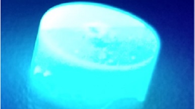
In this study, the researcher from Milano-Bicocca designed a composite material, made of a processable polymeric matrix that incorporates scintillating Metal- Organic Frameworks (MOFs) nanocrystals, thus demonstrating an improvement in the scintillation efficacy. This multicomponent material would allow the fabrication of radiation detector more sensitive and with a better time response to be employed in TOF-PET to enhance the imaging accuracy and scanner sensitivity.