Optics and Optometry
Our research group is dedicated to advancing knowledge in optics, optometry, vision science, and ophthalmic technologies (COMiB – Optics and Optometry Research Center).
Current research projects focus on progressive addition and multifocal lenses, including brain adaptation to these devices, myopia progression, presbyopia, filtering lenses, and the impact of lenses on vision. Additionally, we are developing new optometric and psychometric tests.
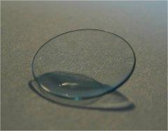
We are also actively engaged in the characterization of contact lens materials, investigating their structure and surface properties (e.g., wettability, tribological analyses, and morphology), corneal analysis, and biomolecular profiling of the tear film. Our precision single-tear profiling approach provides detailed molecular insights, facilitating the identification of disease-specific biomarkers.
The group conducts both fundamental and applied research, integrates clinical optometric practice, and collaborates with industry partners to develop training programs in optics and optometry.
Research Group
Prof. Silvia Tavazzi
Dr. Fabrizio Zeri
Dr. Erika Ponzini
Research Lab
Laboratory – Building U5, Ground Floor, Room T073
IRMA Laboratory – Building U5, First Floor, Room 1078
Optometric clinic – Building U9, First Floor, Rooms 1116-1121
Facilities
The optometry research laboratories are fully equipped. They include phoropters, slit lamps, non-mydriatic retinal cameras with fundus autofluorescence, anterior and posterior segment optical coherence tomographers (OCTs), Scheimpflug tomographers, non-contact tonometers/pachymeters, corneal topographers, closed and open-field ocular aberrometers and autorefractometers, static and kinetic perimeters, keratometers, ophthalmoscopes, retinoscopes, and an eye-tracking and visual psychophysical lab.
In-vitro and ex-vivo research is carried out by UV-visible-NIR spectrophotometry, microvolume spectrophotometry, nanotribometry and other techniques for the characterization of sur-faces, refractometry, instrumentation for photoluminescence and illuminance analyses, fluorescence and polarized optical microscopy, as well as other techniques in collaborations with other research groups.