Laboratory for Crystal Growth and Characterization and Single Crystal X-ray Diffractometry
Scientific Director: Prof. M. Moret
Location: Room 1061, 1st Floor, U5 Building
The CCC-SCXRD laboratory is equipped for the growth and morphological and structural characterization of single crystals of inorganic and organic compounds. It is possible to study in situ by optical microscopy the growth of crystals or their transformation (e.g. mediated by dissolution and Ostwald ripening) and processes due to polymorphic phase transitions. The structural characterization includes the acquisition of X-ray diffraction data (including non-room temperature) on single crystals or microcrystalline powders and the subsequent resolution of the crystalline structure (single crystals only). The instrumentation is completed by the presence of thermostatic systems for crystal growth from solution, by vapor phase transport or by sublimation. Temperature control is available on optical microscopes by means of a heating/freezing stage from approx. -180 to 600 °C.
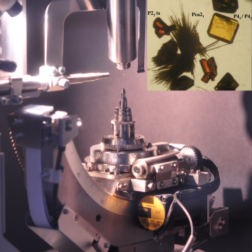
Single crystal X-ray diffractometer
Model: RAPID II (Rigaku)
Technical specifications
- Curved image plate detector 460 x 256 mm2 with 100 μm pixels, photomultiplier for reading the diffracted intensities, χ and φ goniometric movements, X-ray tube with molybdenum anticathode, SHINE monochromator to increase the X-ray flux, digital camera for optical centering of the sample, personal computer for instrument control and data processing;
- Oxford Cryosystems N700 Plus cryostat for sample temperature control from 80 to 500 K;
- CrystalClear instrument management software;
- CrystalStructure data reduction, resolution and structural refinement software;
- RD powder diffraction data acquisition and processing.
The acquisition of the diffraction pattern of organic single crystals and inorganic is carried out in order to:
- determine the unit cell parameters and the space group;
- obtain the atomic coordinates in the solid through the resolution of the structure.
Metallographic Microscope
Model: BX51 (Olympus)
Non-inverted metallographic microscope equipped with polarizer and analyzer for observations in transmitted and reflected light, in bright or dark field.
The microscope is equipped with:
- semi-apochromatic objectives with parallel optical beam (5-10-20-50 magnifications) coupled to the trinocular tube with maximum magnification of 500 X;
- sample holder stage with rotation measurable with goniometer and vernier plus x-y translations also equipped with vernier;
- insert for observation with differential interferential contrast (DIC) according to Nomarski for images with high contrast and vertical resolution;
- possibility of time lapse acquisition of images (even for days).
The metallographic microscope allows:
- the observation of samples and acquisition of images in transmitted and reflected light, with polarized light or with differential contrast according to Nomarski;
- the study of samples as a function of temperature and time (phase transitions, thermal stability, thermal cycles);
- the determination of the size and number of objects displayed through analysis with dedicated software.
Research Stereo-Microscopes
Model: SZX12 (Olympus)
Stereo-microscope equipped with polarizer and analyzer for both transmitted and reflected light observations, in bright or dark field, with oblique illumination, with built-in iris diaphragm for depth of field control. The microscope is equipped with:
- 1X parfocal objective with parallel optical beam coupled to the 10X objective of the trinocular tube with variable magnification from 7 to 90 X (zoom ratio 1:12.86);
- diameter of the field of view 31.4-2.4 mm;
- working distance 70 mm; the microscope is equipped with a long column for the observation of very thick samples (up to about 30 cm);
- diascopic base with sample holder stage with rotation measurable with goniometer and vernier plus x-y translations also equipped with vernier;
- additional lens for magnifications up to 225X;
- possibility of time lapse image acquisition (even for days).
Model: SZX16 (Olympus)
Stereo-microscope equipped with polarizer and analyzer for both transmitted and reflected light observations, in bright or dark field, with oblique illumination, with built-in iris diaphragm for depth of field control. The microscope is equipped with:
- 0.5X and 1X parfocal objectives with parallel optical beam coupled to the 10X objective of the trinocular tube with variable magnification from 7 to 115 X (zoom ratio 1:16.43);
- diameter of the field of view 60-4 mm;
- working distance 70 mm;
- diascopic base with LED lighting and sample holder stage with rotation measurable with goniometer and vernier plus x-y translations also equipped with vernier;
- possibility of timed image acquisition (even for days).
Also available:
- double gooseneck lighting with intensity and color temperature adjustment;
- LED ring lighting.
Heating/freezing Stage
Model: THMS600 (Linkam)
It allows the study of solid or liquid samples in a controlled environment at a temperature from -196°C to +600°C and possible inert gas atmosphere for observation under the optical microscope. This system is equipped with:
- manual x-y translations up to 16 mm for positioning the sample within the area observable under reflection or transmission;
- temperature control from -196°C to +600 °C within 0.1 °C with the possibility of programming temperature ramps from 0.01 °C/min to 130 °C/min; it is possible to program heat treatment cycles using the instrument management program.
The hot stage system can be mounted on the BX51 metallographic microscope or on the SZX series stereomicroscopes using time lapse as well as manual image acquisition.
The hot stage system allows the study of the thermal stability of the samples, of phase transitions, determination of the melting or decomposition point, controlled crystallization from solution using a special fused silica crucible.